Oral airways and bite blocks may both be placed in the mouth during respiratory care, but they are designed to address very different airway problems. In mechanically ventilated patients, the concern is not always upper airway obstruction. In many cases, the more immediate issue is protecting the endotracheal tube from compression caused by jaw clenching, chewing motion, or intermittent biting. When that happens, airflow resistance can change quickly enough to interfere with ventilation delivery, suction access, and airway monitoring.
Clinicians commonly compare oral airways and Bite Blocks because both can appear relevant when an intubated patient has oral movement or tube-related risk. Oral airways are mainly used to maintain a patent upper airway by preventing tongue-related obstruction in unconscious or deeply sedated patients. Bite Blocks are used when the clinical priority is to protect the endotracheal tube from compression. Choosing between them requires a clear understanding of airway anatomy, sedation level, tube position, patient tolerance, suction access, and the specific risk affecting ventilation.
Why Device Selection Matters for ET Tube Protection
Endotracheal tube protection is not the same as upper airway positioning. Once a patient is intubated, the ET tube becomes the controlled pathway for ventilation, oxygen delivery, suctioning, and airway access. If the tube is compressed by biting, the problem occurs inside the artificial airway rather than behind the tongue. A device that improves upper airway patency may not fully address that risk if it does not keep jaw force away from the tube itself.
This difference affects bedside decision-making. A patient with tongue-related obstruction during bag-mask ventilation may benefit from an oral airway because the goal is to keep soft tissue from closing the upper airway. A mechanically ventilated patient who is clenching around an ET tube requires a different type of protection because the concern is tube lumen narrowing, suction difficulty, ventilator pressure changes, and possible tube deformation. The best device is the one that matches the source of airway compromise.
This comparison also matters because device misuse can create new problems. An oral airway placed in a patient who is becoming more awake may trigger gagging, coughing, or agitation. A bite block placed without attention to oral anatomy may create pressure points or limit visualization. Strong airway management depends on choosing a device that solves the immediate clinical problem while preserving access for suctioning, oral care, securement checks, and ongoing reassessment.
How Oral Airways Support Upper Airway Patency
An oral airway is designed to move the tongue away from the posterior pharynx so airflow can pass more easily through the upper airway. It is commonly used during anesthesia, emergency airway management, bag-mask ventilation, and deep sedation when soft tissue collapse is the primary concern. Its structure helps maintain an open passage through the mouth and pharynx in patients who cannot reliably maintain their own airway position.
That function is useful, but it is different from protecting an endotracheal tube. In an intubated patient, the ventilator is not relying on the natural upper airway in the same way because the ET tube is already providing the airway route. An oral airway may improve oral spacing or positioning in some situations, but its main design purpose remains upper airway patency rather than direct ET tube protection. If the patient bites around the tube or applies strong jaw pressure, the oral airway may not consistently prevent compression of the tube lumen.
Tolerance is another major factor. Oral airways are generally best tolerated in unconscious or deeply sedated patients. As protective reflexes return, the device may stimulate gagging, coughing, biting, or agitation. This is why clinicians reassess the device as sedation level changes. A device that is appropriate during deep sedation may become poorly tolerated during emergence, neurological recovery, or ventilator weaning.
For readers exploring the dental use of bite blocks, Does Everyone Get Bite Blocks with Braces? explains how orthodontic bite blocks are used differently from respiratory airway devices used during ventilation.
How Bite Blocks Protect the Endotracheal Tube Differently
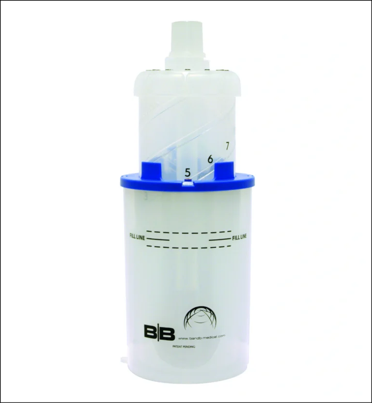
Bite Blocks are designed around a different mechanical goal. Instead of lifting the tongue away from the posterior airway, they create separation between the upper and lower teeth so jaw force is less likely to close directly onto the endotracheal tube. This makes them more specific to tube protection when biting or clenching is the concern. The clinical value comes from reducing direct tooth-to-tube compression rather than improving natural upper airway patency.
This difference becomes important when ventilator performance depends on a consistent tube lumen. If jaw pressure narrows the ET tube, airflow resistance can rise, suction catheters may pass less easily, and ventilator readings may become harder to interpret. Bite Blocks help preserve the shape and access of the tube during periods of oral movement, especially when patients are partially awake, neurologically reactive, or moving through sedation changes.
They also change how oral pressure is distributed. Instead of allowing the tube to absorb repeated biting force, a properly placed bite block helps create a protective spacing point within the mouth. Clinicians still need to monitor lips, tongue, gums, teeth, and mucosa, but the device is selected because the risk is tube compression, not tongue collapse. That is the main functional difference between the two device categories.
To better understand the dental meaning of bite blocks compared with airway-management devices, the educational guide What Is Bite Block & How Does It Help With Braces? provides additional context.
Why Oral Airways May Not Fully Prevent Tube Compression
Oral airways may appear protective because they occupy space in the mouth, but their shape and purpose do not always control where biting force is directed. Depending on the oral airway design, ET tube position, patient dentition, and jaw movement, the patient may still compress the tube against teeth, oral tissue, or the device itself. This is especially relevant when clenching is forceful, repetitive, or related to neurological activity.
Another limitation is that oral airways are not always positioned around the ET tube in a way that protects the tube lumen. The device may maintain tongue position while the ET tube remains vulnerable to lateral movement or direct compression. In real bedside conditions, the problem is rarely just whether a device is present in the mouth. The more important question is whether it protects the actual structure at risk.
This is where device intent matters. Oral airways are selected when the care team needs upper airway support. Bite blocks are selected when the care team needs a protective barrier against biting. Treating them as interchangeable can weaken airway management because it overlooks the difference between soft tissue obstruction and artificial airway compression.
When Clinicians Choose Bite Blocks Over Oral Airways
Clinicians are more likely to choose Bite Blocks when the endotracheal tube is at risk from jaw clenching, chewing motion, intermittent biting, seizure-related activity, agitation, or partial wakefulness. In these cases, the patient already has an artificial airway in place, so the clinical priority is not opening the upper airway around the tongue. The priority is preserving the ET tube pathway so ventilation, suctioning, and monitoring remain more reliable.
The choice may also depend on how the patient is progressing through care. During deep sedation, an oral airway may still have a role if upper airway positioning or oral access is part of the management plan. During lighter sedation or weaning, the same device may become less tolerated because gag reflexes and airway reflexes begin returning. A bite block may be more relevant when the patient still needs the ET tube but has enough jaw activity to threaten tube patency.
Clinical judgment remains central because neither device is selected in isolation. The care team must consider tube depth, securement, oral anatomy, secretion load, risk of aspiration, mucosal condition, sedation strategy, and extubation readiness. The better choice is not based on the device name alone. It is based on whether the airway problem is tongue obstruction, tube compression, or a combination of risks that requires closer reassessment.
Can Oral Airways and Bite Blocks Be Used Together?
In some clinical situations, teams may consider more than one oral airway support approach, but combined use depends on patient anatomy, device compatibility, tube position, oral space, and facility protocol. The primary issue is to avoid crowding in the mouth. Excessive oral equipment decreases visualization, limits access for suctioning, increases pressure on mucosal tissue, and makes securement checks more difficult.
If both upper airway positioning and tube protection are concerns, clinicians must assess which risk is most immediate and which device combination can be used safely without interfering with ventilation management. The mouth is already occupied by the ET tube, securement materials, secretions, and sometimes edema or tongue swelling. Adding devices without enough space or reassessment can create pressure points or reduce access to the tube.
This is why the decision should remain patient-specific. Some patients may need tube protection only. Others may need support for oral positioning, suctioning access, or airway stability during a short procedural window. The safest approach is to use the fewest devices needed to achieve the airway goal while keeping the tube visible, accessible, and regularly reassessed.
Workflow Considerations for Suctioning, Oral Care, and Monitoring
Device selection affects more than tube protection. It also affects how easily clinicians can suction secretions, examine oral tissue, check tube position, adjust securement, and provide oral hygiene. A device that protects one part of the airway setup but limits access to another can create new workflow challenges, especially during longer periods of ventilation when oral care, secretion management, and pressure checks are repeated as bedside procedures.
Suctioning access is a major consideration. If a device crowds the mouth or shifts against the ET tube, it may become harder to pass suction catheters or maintain a clear view of the tube. Oral care access also matters because secretions, dryness, edema, and pressure exposure can change tissue condition over time. Clinicians need enough visibility to identify irritation, mucosal injury, lip pressure, tongue swelling, and device migration before those issues progress.
Ventilator monitoring is also part of the workflow. If tube compression is reduced, pressure and volume changes may become easier to interpret because one source of intermittent resistance has been controlled. If airway alarms continue despite tube protection, clinicians can focus assessment on secretions, bronchospasm, circuit issues, lung mechanics, or other causes. A well-chosen device supports both physical airway protection and clearer clinical troubleshooting.
For clinicians interested in broader respiratory-support workflows, How Bubble CPAP works? explains how airway support strategies differ between invasive ventilation and non-invasive neonatal respiratory support systems.
B&B Medical Technologies Airway Support for Mechanical Ventilation Workflows
B&B Medical Technologies develops respiratory and airway-management products designed around practical bedside workflows in critical care and neonatal environments. The company focuses on solutions that help clinicians support airway stability, respiratory access, secretion management, ventilation efficiency, and safer respiratory workflows during patient care.
Its airway-support portfolio is built for real clinical conditions where device selection, tube movement, oral access, secretion burden, patient agitation, and airway reassessment all affect ventilation management. Products designed for respiratory care must work within fast-moving bedside environments where clinicians need dependable support without creating additional complexity. B&B Medical Technologies approaches airway support with that workflow reality in mind.
Why Bite Blocks Are Part of Comprehensive Airway Management
Bite Blocks should be viewed as one part of a broader airway-management plan rather than a replacement for clinical assessment. Mechanical ventilation requires coordinated attention to tube securement, cuff management, ventilator synchrony, secretion clearance, humidification, oral hygiene, sedation strategy, neurological status, and extubation readiness. If tube biting occurs repeatedly, clinicians may also need to evaluate discomfort, delirium, pain control, neurological stimulation, or worsening respiratory intolerance.
Ongoing review remains central because the airway needs change throughout the course of ventilation. For example, in deeply sedated patients, the priorities may be managing secretions, tube position, and stable ventilation, while during the weaning or neurologic recovery phases, clinicians may be more concerned with spontaneous breathing effort, return of protective reflexes, and readiness for extubation. Bite Blocks can be helpful in maintaining tube patency when biting activity may threaten ventilation, but should always be used with ongoing clinical assessment.
Frequently Asked Questions
An oral airway helps maintain upper airway openness by reducing tongue-related obstruction. Bite Blocks protect the endotracheal tube from being compressed by biting or jaw force during ventilation.
Some oral airways may provide only slight separation between the teeth and tube, but are not designed to redirect the biting force away from the ET tube. Patients with strong jaw activity may still be able to compress the tube.
They help prevent biting and restriction of the endotracheal tube that may help maintain airflow, suctioning access, and reduce ventilator instability during care.
Yes. Ongoing or aggressive biting can compress the inside of the tube enough to affect airflow, alter ventilator measurements, and occasionally require tube adjustment or exchange.
They may be considered together only when clinically appropriate and compatible with the patient’s oral anatomy, tube position, and airway goals. Clinicians must avoid crowding, pressure injury, reduced visualization, and limited suctioning access.
No. The devices serve different airway purposes. Oral airways are used primarily for the maintenance of upper airway patency, and Bite Blocks are used to minimize endotracheal tube compression.
Clinicians should assess oral tissue condition, tube stability, secretions, device placement, suction access, and ventilation effectiveness throughout care.
Any oral device may create pressure if poorly positioned, left in place too long without reassessment, or used in patients with fragile tissue. Regular oral assessment helps reduce that risk.